Who are you, and what do you do?
My name is Cathrina Geldard (@CathrinaGeldard on Twitter), and I live in Hobart, Tasmania, Australia. I am a trained naturopath, currently working as a research assistant and PhD candidate at the National Centre for Naturopathic Medicine at Southern Cross University.
My work looks at how herbal medicines used by traditional healers can help treat chronic infections that have become resistant to modern antibiotics. My Masters research looked at the antibiotic effects of medicinal mushrooms that had been shown to be cytotoxic (cancer-fighting) and to improve immune function, and now in my PhD I am focusing on medicinal plants from the Balkan Peninsula for fighting urinary tract infections (UTIs) - the most common bacterial infection in humans, which can sometimes effect people for decades.
Outside of work and study, my time is spent volunteering with Herbalists Without Borders; hanging out with my husband, adult children, and pets; doing home renovations; and exploring the beautiful state of Tasmania I live in.
What hardware do you use?
My work is split between lab work and non. In the lab my basic tools are pipettes and tips, test tubes, petri dishes, growth mediums, and inoculation loops. When necessary, items are sterilised in an autoclave before use.
The largest part of my work in the lab is growing tough little bacterial colonies called "biofilms" in conditions similar to that in a human bladder. Though colonies that cause infections in humans are thought to be often made up of multiple bacteria - usually E. coli and friends - it turns out they're very stubborn about teaming up in lab conditions, so I spend a lot of my time trying to make them stop killing or avoiding each other.
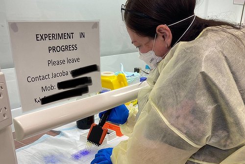
I grow my static biofilms on Merck polystyrene 96-well plates. I mainly use the non-tissue culture ones because they are significantly cheaper, and I have found in my experiments there is little difference when I use the more expensive tissue culture plates.
After growing the biofilms in an incubator and staining them purple, the biofilm biomass is measured by reading absorbance at the 595-nanometre wavelength with an iMark Microplate Absorbance Reader. This tells me if the biofilm colony grew and, if so, how well.
To more closely mimic the bladder environment where these bacteria thrive, I grow the biofilms on actual bladder cells. We then place these in "flow cells": a setup where the cell growth area is attached to a set of pumps that continuously pass liquid through it. This allows direct microscopic examination of the biofilms as they develop and means the growth environment can be carefully controlled and easily changed. We use the Transmission Flow Cell FC 285-AL. This model has injection ports where you can add bacteria and inject herbal medicine treatments during the experiment. It has a maximum flow rate of 3.5mL/min which is enough to study biofilms in the similar conditions to the bladder - where they have to attach to and bury into the wall cells to avoid getting flushed away.
After growing the biofilms in the flow cell, I look at them under the microscope and produce images of the biofilm. We have been using a Motic B3 series microscope. With this microscope I can only see there is a biofilm and quantify how much of it there is, but I need more detail than that so we are upgrading to a ZEISS Apotome 3. With this microscope, I'll be able to see the biofilm's 3D structure, assess its structural parameters, identify the proportion of living and dead cells within the biofilm, and more.
Any herbal medicines that show success at fighting biofilms will be classified and characterised by mass spectrometry using the Thermo Scientific ISQ EC Single Quadrupole Mass Spectrometer. Mass spectrometry is an analytical technique that uses ionized vapours to identify and quantify compounds within a sample such as a herbal medicine preparation, and elucidate the structure and chemical properties of different molecules within. The method plays an important role in determining the specific "bioactive" compounds which are associated with any beneficial activity we observe in our experiments.
Outside of the lab, research mostly involves long days of reading, analysing data and writing. When you start a PhD at SCU, you are loaned a computer by the university. Mine is a Dell Latitude 5420 with an Intel i5 processor. I have only been using this for 4 months and it's fine.
Previously I had a hand-me-down 2017 13" MacBook Pro laptop from my son-in-law but my university had no technical support for Mac users and I knew I'd have to use a bunch of new software for my research, so I swapped back to the Windows one they gave me. I miss the MacBook mostly because I loved having my phone (a second-hand iPhone 11 Pro) synced with the computer so I could receive texts while I work. As a mum of three young adults, I always have one eye on my text messages.
And what software?
My university computer runs on Windows 10, so most of the tools I use are whatever Microsoft provides or the university endorses.
- For communication, I use email or Microsoft Teams - though my supervisor and I are communicating through WhatsApp right now while she is overseas.
- For written work, I mainly use Microsoft Word 365 but I have just recently started learning LaTeX via Overleaf. Now that I have gotten to the part of my PhD where I will be submitting more papers to scientific journals for publication, my daughter (who is also a doing a PhD) has convinced me that using LaTeX will reduce my workload when the same paper needs reformatting for submission to different journals.
- For posters and presentations, I use Microsoft PowerPoint 365 - although when I had my Mac I preferred Keynote.
- For data analysis, I am currently using Microsoft Excel 365, NVivo 12 and IBM SPSS.
- For managing citations, I use Zotero 6.0.8.
- For research surveys, such as collecting information about traditional medicine use from practitioners, I use the Qualtrics survey platform.
But most of my time is spent finding and reading research to keep up with developments in my field, so the software I use the most by far is Google Chrome. Then I back up all of my work in Microsoft OneDrive and Google Drive.
What would be your dream setup?
If my research budget was limitless, I would love to use the latest organoid or organ-on-a chip technology to study urinary tract infections, as this could better capture the complexity of human host physiology and how that impacts bacterial growth.
Bladder organoids (essentially tiny bladders grown from stem cells) allow study of the cell interactions between uropathogens and bladder cells, including factors introduced by the multi-layered structure of the bladder wall. Horsley et al. (2018) developed an organoid from human progenitor cells which demonstrates key structural hallmarks and biomarkers of the urothelium (bladder lining). With this, they showed how when bacteria invade the bladder cells, the urothelium resorts to sloughing off its outermost cells to try to get rid of the infection.
The bladder-on-a-chip technology is slightly simpler, like a 3D printed circuit of cells that grow together under conditions that closely mimic the bladder–including a simulated urine flow and mechanical forces to simulate the expansion and contraction the bladder experiences as it fills and empties of urine. Sharma et al. (2021a, b) developed bladder chip technology used alongside bladder organoids (although these organoids were from mice, which I don't want to do). With these, they captured in great detail an E. coli invasion of urothelial cells, their persistence to antibiotics, and the dynamics of immune cell responses to infection.